How do scientists make a karyotype?
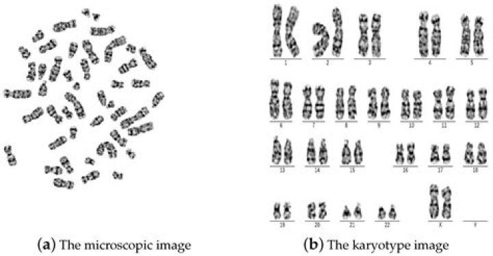
A karyotype is simply a picture of a person’s chromosomes. In order to get this picture, the chromosomes are isolated, stained, and examined under the microscope. A picture of the chromosomes is taken through the microscope. Then, the picture of the chromosomes is cut up and rearranged by the chromosome’s size.
How is a karyotype created and arranged?
In a human karyotype, autosomes or “body chromosomes” (all of the non–sex chromosomes) are generally organized in approximate order of size from largest (chromosome 1) to smallest (chromosome 22). Using this naming system, locations on chromosomes can be described consistently in the scientific literature.
What are the three steps scientists use to make a karyotype?
Let’s take a look at these steps so you can understand what is happening during the time you are waiting for the test.
- Sample Collection.
- Transport to the Laboratory.
- Separating the Cells.
- Growing Cells.
- Synchronizing Cells.
- Releasing the Chromosomes From Their Cells.
- Staining the Chromosomes.
- Analysis.
What will a karyotype show you?
Karyotype is a test to identify and evaluate the size, shape, and number of chromosomes in a sample of body cells. Extra or missing chromosomes, or abnormal positions of chromosome pieces, can cause problems with a person’s growth, development, and body functions.
Why do scientists need to prepare a karyotype?
Chromosomes are packets of genetic information, and the shape, size, and number of chromosomes an individual has can help scientists look for genetic disorders. A karyotype is essentially a picture of an organism’s chromosomes. It allows scientists to visually examine the chromosomes. There are a few steps to follow to properly prepare a karyotype.
How does a karyotype take a picture of a chromosome?
A karyotype is a representation of the person’s chromosomes. To get a picture of chromosomes, the chromosomes are isolated, stained, and checked under the microscope. Through the microscopic examination, a picture of the chromosomes is taken, cut up, and rearranged according to their size from largest to smallest.
How are dividing cells harvested in karyotyping?
Karyotyping involves analysis of the entire chromosome complement through the microscope. Dividing cells are harvested during metaphase, the time of greatest chromosome condensation, by disruption of the spindle using drugs such as colchicine.
What’s the difference between karyotyping and karyogram?
Karyotyping is a technique to grow chromosomes, while karyotype is a method or procedure to arrange it using either manual method or computational software. “A process to pair and arrange chromosomes in order to encounter any defect is known as karyotype or karyogram.”